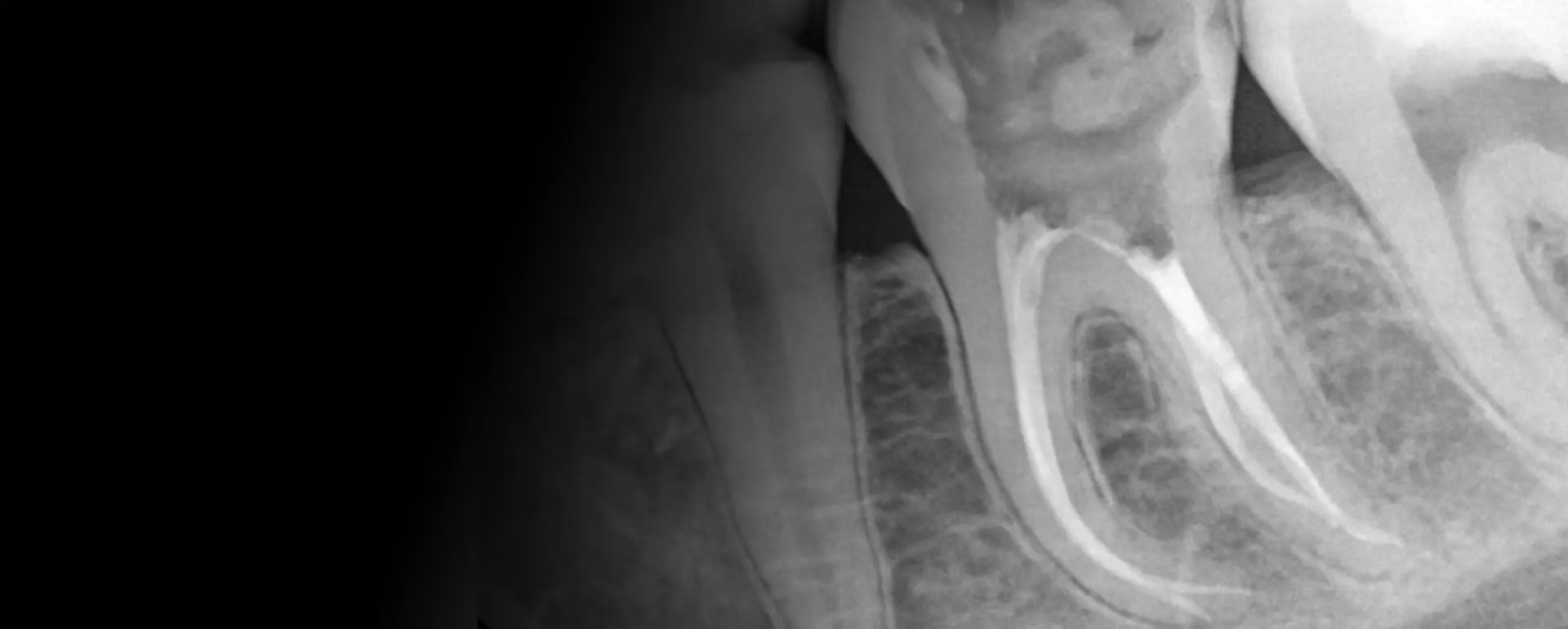
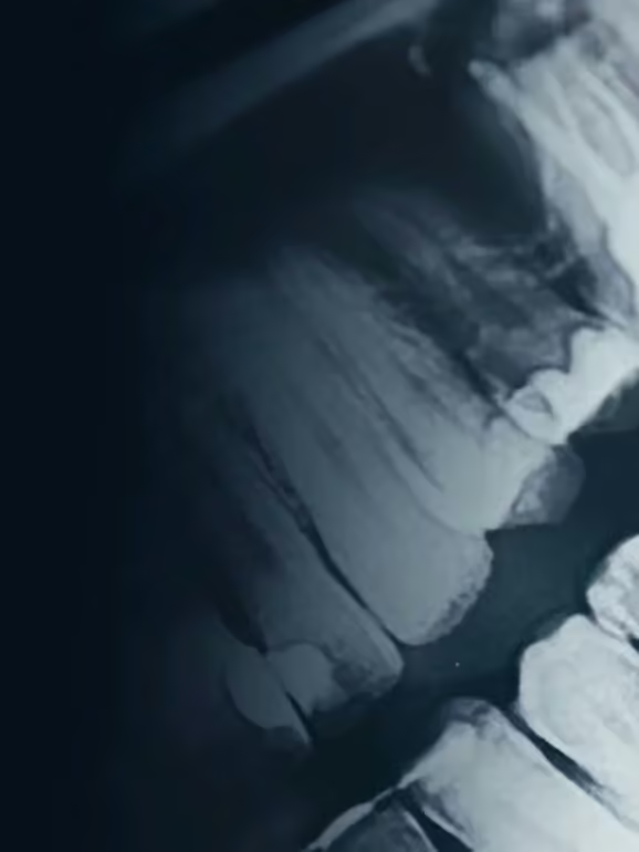
Bone loss is not the price you pay for root canal treatment. It is the other way round: the very aim of this treatment is to stop the process that destroys the bone. If the bone defect grows despite treatment, it is a sign that the infection has not been brought under control.
This article explains where bone loss around the root comes from and how to recognise the problem. It then describes what a radicular cyst is and what treatment options remain before tooth extraction comes into play.
Bone loss after root canal treatment: when is it a complication?
Loss of bone tissue after root canal treatment results from chronic inflammation and bacterial infection, not from the removal of the pulp as such. Bacteria in the root canal can cause chronic inflammation and bone loss, because in response the body resorbs its own bone tissue around the root tip. Incompletely treated infection therefore leads directly to bone loss.
Properly performed treatment works in exactly the opposite way. It removes the source of infection, so bone loss can reverse after successful root canal or surgical treatment.
It is also worth dispelling the myth that a “dead” tooth destroys bone. The dental pulp is indeed removed, but the tooth remains in its socket, held by the living fibres of the periodontal ligament, and when you chew it stimulates the bone mechanically. The root cementum and the periodontal ligament remain alive. Losing a tooth after root canal treatment, on the other hand, removes that mechanical stimulation, and only then does bone loss follow.
Causes of bone loss around the tooth root
The mechanism is almost always the same: bacteria have survived somewhere. Only the route by which they got there differs, and the most common complications of root canal treatment come down to exactly this.
Chronic periapical inflammation
Chronic inflammation around the root tip leads to bone loss and is the main cause of this complication. Inflammation of the periapical tissues is most often caused by bacterial infection, which frequently starts from untreated, advanced tooth decay.
Chronic periapical inflammation can last for years without symptoms. The patient feels nothing, while the lesion around the root tip gradually grows, visible only on an X-ray.
Incompletely filled canals and leaking restorations
Any empty space left in the canal becomes a reservoir for bacteria. This happens with unusual anatomy, when an extra canal goes undetected, or with narrow and sharply curved canals.
The second route of reinfection is the crown. A leaking crown or filling increases the risk of reinfection and bone loss, because bacteria from the mouth get back inside the tooth despite correctly filled canals. Irritation from sealer material or gutta-percha pushed out through the apical opening beyond the root also plays a part.
Vertical root fracture and overloading
A vertical fracture creates a permanent gap connecting the mouth with the jawbone. Bone loss then progresses along the fracture line, and a standard X-ray usually does not show it. A CBCT scan settles the question.
A separate factor is mechanical overloading of the tooth, which can cause bone loss even without infection. On top of this there is gum disease, which leads to bone loss around the tooth regardless of the condition of the canals. Mechanical injury and past trauma can also be a starting point, as can pulp necrosis that develops years after the event.
Periapical periodontitis: symptoms and treatment
The symptoms are intense toothache and swelling of the tissues, but this applies to the acute form. The chronic form can be silent, which is why diagnosis relies on the X-ray image.
- pain when biting and on tapping, indicating a reaction in the periodontal ligament,
- a sinus tract (fistula) on the gum or recurring swelling around the affected tooth,
- a dark area (radiolucency) at the root tip visible on an X-ray,
- tooth mobility, as a tooth that has had root canal treatment can become loose because of bone loss.
Treating inflammation of the periapical tissues requires endodontic treatment, that is, removing the infection from inside the tooth. Periapical lesions take between 6 and 24 months to heal, so assessing whether treatment has worked calls for patience and follow-up X-rays.
Radicular cyst: causes and types of cysts
A radicular cyst develops as a result of tooth necrosis, around a non-vital tooth, as a consequence of long-standing inflammation. It has its own capsule and a fluid-filled cavity, and a radicular cyst causes resorption of the surrounding bone.
On an X-ray, a dental cyst appears as a smooth, clearly defined dark area. As it grows, the lesion displaces the roots of neighbouring teeth and may press on nearby anatomical structures, including the floor of the maxillary sinus.
Types of cysts seen in dental practice
In dental practice, the most commonly diagnosed cyst is the radicular cyst, but it is not the only form.
- radicular cyst, forming at the tip of a tooth with dead pulp,
- dentigerous cyst, developing around the crown of an unerupted tooth,
- dentigerous cysts around impacted wisdom teeth, usually discovered by chance,
- residual cyst, remaining after the causative tooth has been extracted without the lesion being removed.
A dental cyst can be the result of inadequate root canal treatment, when infection persists despite the canals having been filled. The causative tooth then becomes a source of infection that has to be removed or re-treated.
Treatment of a radicular cyst
Treating a dental cyst often requires surgical removal of the lesion together with its capsule. Small cysts, however, are sometimes treated conservatively: after effective disinfection of the canals, the lesion regresses and the bone rebuilds.
Surgical removal of the lesion is usually combined with root-end resection (apicoectomy). An untreated radicular cyst can lead to deformity of the jawbones, so putting off a decision has a real cost here.
Worrying symptoms in the mouth and beyond
Some warning signs do not concern the tooth itself, and that is exactly why they tend to be ignored. Untreated inflammation can lead to serious complications, including systemic ones.
- severe pain that does not ease with painkillers,
- swollen lymph nodes on the side of the affected tooth,
- feeling unwell, a low-grade fever, general weakness,
- a feeling of fullness and pressure in the gum tissue around the lesion,
- facial swelling and difficulty opening the mouth.
In advanced cases the infection spreads to the surrounding tissues, and in extreme cases involvement of the maxillary sinus and even inflammation of the cranial nerves have been described. Such situations are rare, but they show that inflammation of the gums and the tissues around the root is not a purely local problem. It has a direct bearing on the health of the whole body.
When is antibiotic treatment needed?
Antibiotics may be recommended when symptoms are severe, or when there is swelling, fever or spreading infection. They do not, however, replace treatment of the underlying cause.
Antibiotics calm the symptoms, but they do not remove bacteria from inside the canals, because the drug does not reach dead tissue that has no blood supply. Without cleaning and shaping the canals or surgical treatment, the problem returns once the medication is stopped. The decision to prescribe it is made by the dentist after a clinical examination.
Root canal re-treatment as the first step
When the original treatment has failed, the first option considered is root canal re-treatment. The procedure involves removing the old filling, cleaning and disinfecting the entire root canal system again and sealing it tightly once more.
We carry it out under a ZEISS Extaro 300 microscope, because removing old material and finding missed canals without magnification can be impossible. Root canal treatment may be split over several visits, especially when exudate persists in the canals.
Root canal re-treatment of a single-canal tooth costs 2000 PLN in our price list, and of a multi-canal tooth 3000 PLN. Removing a broken instrument from a canal is priced separately at 1000 PLN. A successful procedure removes the source of infection, and the body then rebuilds the bone by itself.
Root-end resection (apicoectomy) and surgical endodontic treatment
When root canal re-treatment is not possible or has not worked, surgical endodontic treatment remains. The most common procedure of this kind is root-end resection (apicoectomy), in which the infected tip of the root is removed together with the inflammatory lesion and the canal is sealed from the cut end.
One indication can be a large post and core, whose removal would risk fracturing the root. In the Flow Dental Studio price list, root-end resection costs 3500 PLN for a front tooth and 4000 PLN for a back tooth.
In some cases the procedure is combined with filling the bone defect. Bone regeneration procedures can support the rebuilding of lost bone, especially where lesions are large.
Rebuilding bone: guided regeneration
Where the defect is extensive, guided bone regeneration is used. A bone substitute material is placed where the lesion was removed, acting as a scaffold, and the whole area is covered with a membrane that prevents soft tissue from growing in.
Rebuilding a bone defect corresponds to the bone grafting (regeneration) item in our price list, priced from 2500 PLN. The final cost depends on the size of the lesion and the type of material used.
The bone itself takes months to rebuild. Healing after large defects is sometimes only assessed after a year or two, which is why follow-up X-rays are scheduled every 6 to 12 months.
Tooth extraction: when is it the last resort?
Extraction is considered only when other methods have failed. Removing the tooth can be the only option in a few situations.
- a vertical root fracture running along its whole length,
- tissue destruction that makes a tight, sealed restoration impossible,
- extensive bone loss causing the tooth to become loose,
- recurring infection despite re-treatment and surgery.
This is exactly how teeth are lost, because the bone defect weakens the tooth's stability. After extraction, replacement of the missing tooth is planned to prevent further bone loss in that area. Removing the tooth therefore does not end treatment; it moves it on to another stage.
How to prevent it? Keeping inflammation under control
What has the greatest influence on the outcome is what happens immediately after root canal treatment and in the years that follow.
- a tightly sealed crown restoration made without delay once treatment is complete,
- regular check-ups with an X-ray, even when there are no symptoms,
- daily oral hygiene, as it limits gum inflammation,
- early treatment of tooth decay, before it reaches the pulp,
- controlling bite overload and bruxism.
An X-ray or CBCT scan is recommended to assess the condition of the bone after root canal treatment, and it is the only objective way to verify it. Absence of pain does not confirm healing, because in most cases a chronic lesion does not hurt. The patient's general health is also important here, as it determines how quickly each patient heals.
What does this mean for you?
Bone loss is a symptom, not a diagnosis. There is always a specific cause behind it: persistent infection, a leaking seal, a fracture or overload. Its cause is established through examination and an X-ray, not guesswork.
The good news is that once the source of inflammation is removed, the bone rebuilds itself. The bad news is that gradual bone loss progresses without symptoms, so without follow-up X-rays no one will catch it in time.